The Heart Study Guide
Introduction
One of the most vital life-sustaining organs is the four-chambered muscular organ, the human heart. It’s the same size as a clenched fist, one of the strongest muscles in the human body, and weighs around 200 to 425 grams.
Structure of the Heart
The human heart is a muscular organ with four chambers. The majority of its volume is inclined to the left side of the sternum. A membranous pericardium surrounds the heart.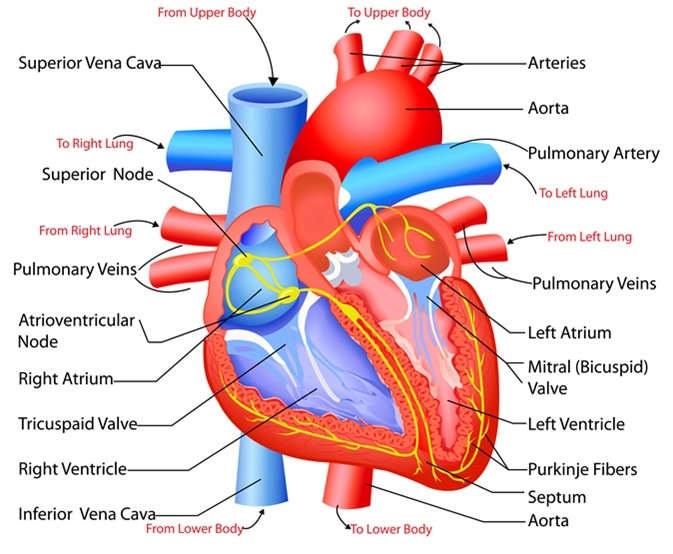 Source
1) Layers of the Heart Wall
The heart is made up of three distinct layers of tissue.
- Endocardium, the innermost layer
- Epicardium, the outermost layer
- Myocardium is a thick layer of specialized muscle between these two layers.
2. Chambers of the Heart
The human heart consists of four chambers:
- Right atrium
- Right ventricle
- Left atrium
- Left ventricle
The atriums are comparatively thin-walled chambers that receive blood from the veins. The ventricles are thick-walled chambers that pump blood away from the heart to all the body parts.
The difference in the thickness of the ventricular wall of the heart is due to the difference in the volume of myocardium present, which is essential as ventricles have to pump blood against greater resistance while maintaining high pressure.
The right atrium gets deoxygenated blood from everywhere across the body through veins. The pulmonary veins bring oxygenated blood from the lungs to the left atrium.
3) Valves of the Heart
The heart has four valves that allow the blood to flow unidirectionally.
- The valve between the atrium and the ventricle is called the atrioventricular valve.
- The semilunar valve is at the base of the large blood vessels that leave the ventricle.
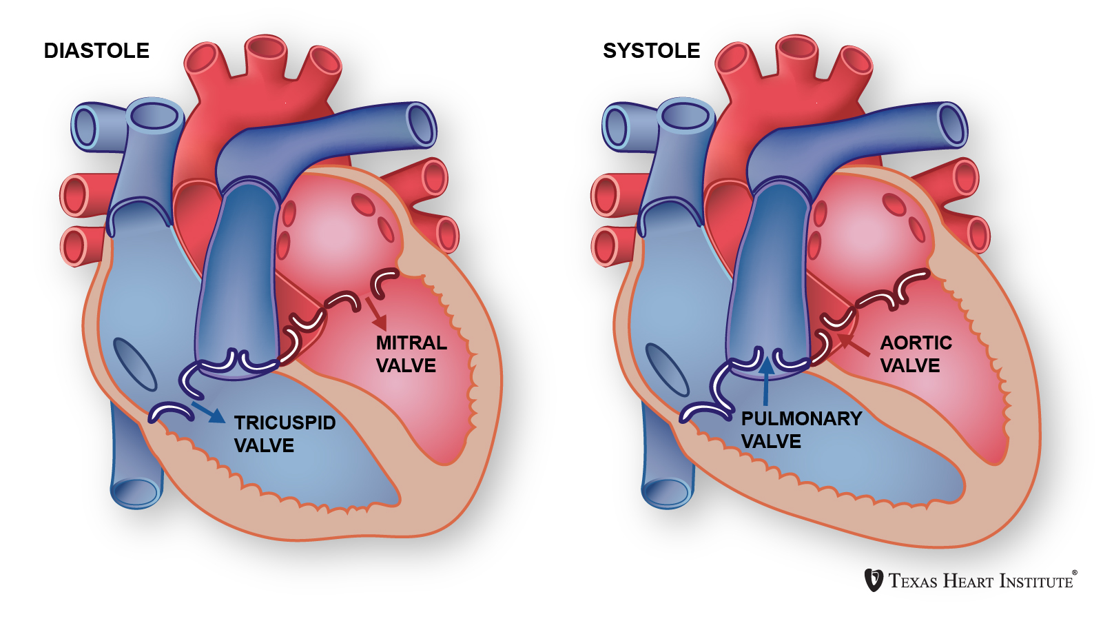
- The right atrioventricular valve is also called the tricuspid valve.
- The left atrioventricular valve is called the bicuspid or mitral valve. It has only two cusps and resembles a miter in structure.
- The right ventricle and pulmonary artery valve are called the pulmonary semilunar valve.
- The valve between the left ventricle and the aorta is the aortic semilunar valve.
- The atrioventricular valves close shut during ventricular contraction, preventing blood from flowing back into the atrium.
- The semilunar valves close when the ventricles relax, preventing blood from flowing back into the ventricles.
4) Pathway of Blood through the heart
- The superior and inferior vena cava and the coronary sinus provide deoxygenated blood to the right atrium.
- The right atrium contracts, forcing blood into the right ventricle through the tricuspid valve. The right ventricle then contracts, allowing blood to travel through the pulmonary artery and the lungs via the pulmonary valve.
- The blood is oxygenated in the lungs before returning to the heart and entering the left atrium through the pulmonary veins.
- The left atrium contracts and forces blood into the left ventricle through the bicuspid valve
- The left ventricle sends oxygenated blood into the aorta via the aortic semilunar valve, circulated throughout the body.
5) Blood Supply to Myocardium
- The heart wall’s myocardium is a functioning muscle that requires a steady supply of oxygen and nutrients to operate properly.
- As a result, the heart muscle contains an extensive network of blood capillaries that transport oxygen to contracting cells and carry waste materials out of the heart musculature.
- Blood enters the system of the heart’s veins after passing via the capillaries of the heart muscle (coronary veins).
- The ascending aorta branches into the right and left coronary arteries, which supply blood to the myocardium’s wall.
- The coronary sinus then carries the deoxygenated blood to the right atrium.
Functions of the Heart
- Pumping oxygenated blood to the body.
- Hormones and other essential compounds are pumped to various body regions by the heart.
- Receiving deoxygenated blood and pumping it to the lungs for oxygenation and transporting metabolic waste products from the body to organs like the liver and kidneys for detoxification and waste removal.
- Maintaining constant blood pressure in the blood vessels.
Conclusion:
- One of the most vital life-sustaining organs is the human heart.
- It’s a four-chambered muscular organ.
- The size of the human heart is the same as a clenched fist.
- The human heart is one of the strongest muscles in the human body.
- Humans adults have 4 functional valves to ensure unidirectional flow of blood.
FAQs:
1. What is a heart made up of?
The heart is made of three layers of tissue. Endocardium, the thin inner lining of the heart chambers that also forms the surface of the valves. The myocardium, the thick middle layer of muscle that allows your heart chambers to contract and relax to pump blood to your body. Pericardium, the sac that surrounds your heart.
2. What is the Human Heart weight?
The average gross weight of the heart was 289.6 g for men at the age of 31-40 years, 284.7 g for women at the same age. At 61-70 years, the average weight was 345.9 g for men and 285.1 g for women.
3. What are the four main functions of the heart?
The four primary functions of the heart are:
- Pumping oxygenated blood to the body.
- Hormones and other essential compounds are pumped to various body regions by the heart.
- Receiving deoxygenated blood and pumping it to the lungs for oxygenation and transporting metabolic waste products from the body to organs like the liver and kidneys for detoxification and waste removal.
- Maintaining constant blood pressure in the blood vessels.
4. Can a person live without a heart?
No. the human heart is the most critical organ of the body to keep a person alive.
5. Can a human have two hearts?
No, a human cannot have two hearts.
6. How does the heart last so long?
The human heart comprises cardiac muscles, which are stronger than normal muscles. The endurance of these muscles is very high, and they do not get fatigued and keep functioning till death.
7. What is the total artificial heart?
A total artificial heart is a surgically implanted pump to replace sick or damaged heart ventricles to supply circulation to the body. This condition is a temporary way to prolong life. Still, the permanent cure is treating the heart completely or a heart transplant as a last resort.
8. Are you dead if your heart stops?
Quite often the heart can stop beating due to various reasons. Most commonly, no heartbeat indicates death, but this is not compulsory that a still heart means death. Instant administration of CPR and defibrillation can restart the heart back to normal functioning. But it is important to note here that immediate administration of CPR is important. After the heart stops beating, there is only a resort of a couple of minutes until permanent neurological damage is not coming back.
We hope you enjoyed studying this lesson and learned something cool about the Human Heart! Join our Discord community to get any questions you may have answered and to engage with other students just like you! Don’t forget to download our App to experience our fun, VR classrooms – we promise, it makes studying much more fun! 😎
Sources:
- Structure of the Heart. https://training.seer.cancer.gov/anatomy/cardiovascular/heart/structure.html. Accessed Nov 26, 2021.
- Heart. https://flexbooks.ck12.org/cbook/ck-12-biology-flexbook-2.0/section/13.24/primary/lesson/heart-bio/. Accessed Nov 26, 2021.
- Your Heart & Circulatory System. https://kidshealth.org/en/kids/heart.html. Accessed Nov 26, 2021.
- Heart disease. https://www.mayoclinic.org/diseases-conditions/heart-disease/symptoms-causes/syc-20353118. Accessed Nov 26, 2021.