CBSE Class 11 Biology Chapter 18 Revision Notes Part 2
Chapter 18: Body Fluids and Circulation
The blood circulatory system, also known as the cardiovascular system, consists of the heart and the blood vessels running through the entire body. It delivers nutrients and oxygen to all cells in the body.
Circulatory Pathways
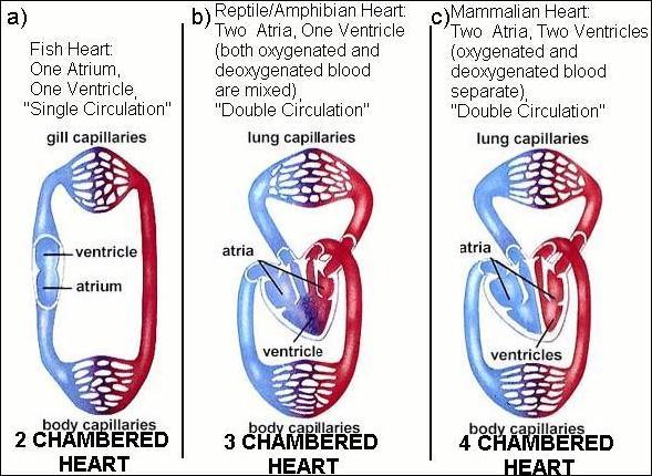
The circulatory patterns are of two types – open or closed.
-
In open circulatory system, blood vessels are absent and blood pumped by the heart passes into open spaces or body cavities called sinuses. Open circulatory system is primarily found in invertebrates such as arthropods and molluscs.
-
In closed circulatory system, the blood pumped by the heart is alwayscirculated through a closed network of blood vessels and the blood does not stay in body cavities. Many vertebrates such as annelids, chordates as well as humans have have a closed circulatory system.
Human Circulatory System
Heart is a mesodermally derived organ, situated in the thoracic cavity, in between the two lungs, slightly tilted to the left. It has the size of a clenched fist. Some facts about the human heart are:
- The heart has four chambers, two relatively small upper chambers called atria and two larger lower chambers called ventricles.
- The opening between the right atrium and the right ventricle is guarded by a valve formed of three muscular flaps or cusps, the tricuspid valve. On the other hand, the opening between the left atrium and the left ventricle is guarded by bicuspid or mitral valves.
- The valves in the heart allows the flow of blood only in one direction, i.e., from the atria to the ventricles and from the ventricles to the pulmonary artery or aorta.
- A patch of the nodal tissue present in the right upper corner of the right atrium is called the sino-atrial node (SAN).
- Another mass of the nodal tissue seen in the lower left corner of the right atrium close to the atrio-ventricular septum is called the atrio-ventricular node (AVN).
- The SAN can generate the maximum number of action potentials, i.e., 70-75 min–1, and is responsible for initiating and maintaining the rhythmic contractile activity of the heart. Therefore, it is called the pacemaker.
Cardiac Cycle
Let’s take a look at how the heart functions.
- At the start of the cycle, all the four chambers of heart are in joint diastole (a relaxed state).
- As the tricuspid and bicuspid valves open, blood flows into the left and the right ventricle.
- The semilunar valves are closed at this stage.
- The SAN generates an action potential which stimulates both the atria.
- The action potential then reaches the AV node, from where the the bundle of His then transmits it to the ventricular musculature.
- This causes the ventricular musclesto contract, (ventricular systole), the atria undergoes relaxation (diastole)
- The ventricular pressure increases causing the closure of tricuspid and bicuspid valves inorder to stop the attempted backflow of blood into the atria.
- This cycle gets repeated again with the decrease in the pressure inside the ventricles.
- This sequential event in the heart which is cyclically repeated is called the cardiac cycle.
- During each cardiac cycle two prominent sounds — lub, which is associated with the closure of the tricuspid and bicuspid valves and dub, which is associated with the closure of the semilunar valves.
Electrocardiograph (ECG)
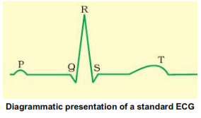
ECG is a graphical representation of the electrical activity of the heart during a cardiac cycle.Each peak in the ECG is identified with a letter from P to T that corresponds to a specific electrical activity of the heart:
- The P-wave represents the electrical excitation (or depolarisation) of the atria, which leads to the contraction of both the atria.
- The QRS complex represents the depolarisation of the ventricles, which initiates the ventricular contraction.
- The contraction starts shortly after Q and marks the beginning of the systole.
- The T-wave represents the return of the ventricles from excited to normal state (repolarisation). The end of the T-wave marks the end of systole.
- The heart beat rate of an individual can be determined by counting the number of QRS complexes that occur in a given time period.
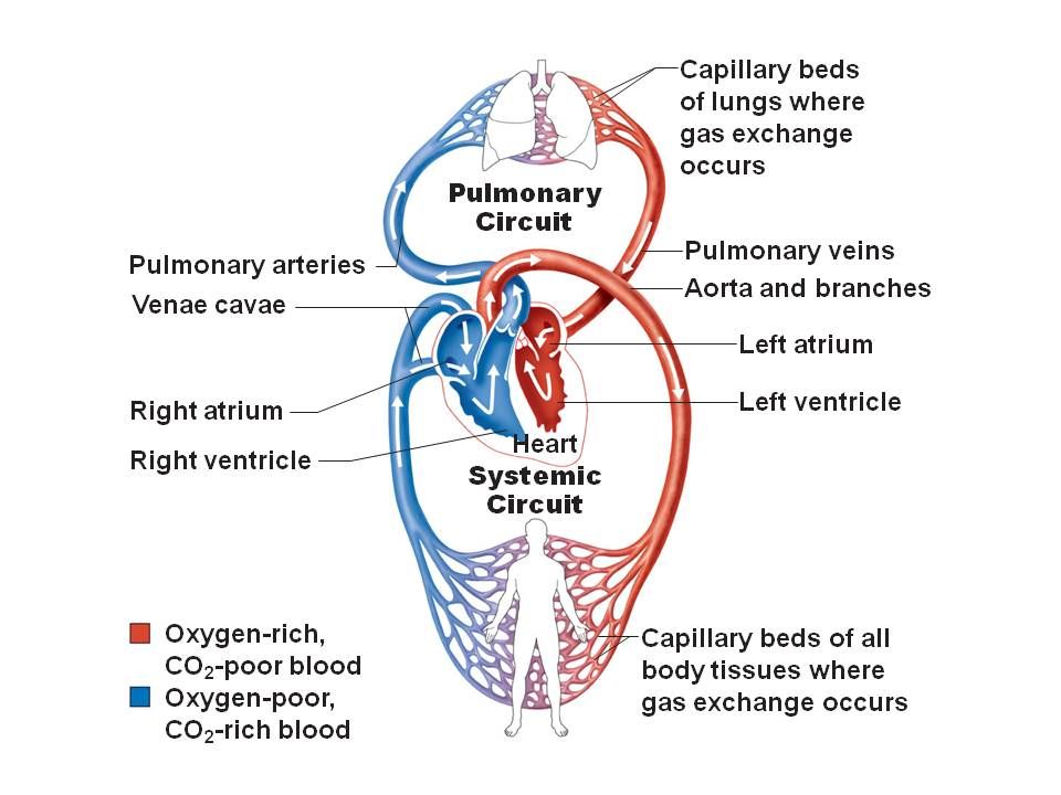
Double Circulation
The deoxygenated blood pumped into the pulmonary artery is passed on to the lungs from where the oxygenated blood is carried by the pulmonary veins into the left atrium. This is called double circulation and it constitutes both pulmonary circulation and systemic circulation.
Systemic circulation: The oxygenated blood entering the aorta iscarried by a network of arteries, arterioles and capillaries to the tissuesfrom where the deoxygenated blood is collected by a system of venules,veins and vena cava and emptied into the right atrium.
Pulmonary circulation: The deoxygenated blood pumped into the pulmonary artery is passed on to the lungs from where the oxygenated blood is carried by the pulmonary veins into the left atrium.
Regulation of Cardiac Activity
- A special neural centre in the medulla oblangata can moderate the cardiac function through autonomic nervous system (ANS).
- Neural signals through the sympathetic nerves can increase the rate of heart beat, the strength of ventricular contraction and thereby the cardiac output.
- On the other hand, parasympathetic neural signals decrease the rate of heart beat.
Disorders of the Circulatory System
- High Blood Pressure (Hypertension): Hypertension is the term for blood pressure that is higher than normal (120/80). High blood pressure leads to heart diseases and also affects vital organs like brain and kidney.
- Coronary Artery Disease (CAD): Coronary Artery Disease, often referred to as atherosclerosis, affects the vessels that supply blood to the heart muscle.
- Angina: Also called ‘angina pectoris’, it is the acute chest pain that appears when enough oxygen does not reach the heart muscle.
- Heart Failure: Heart failure means the state of heart when it is not pumping blood effectively enough to meet the needs of the body.
Sources:
BODY FLUIDS AND CIRCULATION. https://ncert.nic.in/textbook/pdf/kebo118.pdf Accessed 20 Dec, 2021.
]]>