CBSE Class 11 Biology Chapter 20 Revision Notes
Chapter 20: Locomotion and Movement Revision Notes
Movement accounts for various critical tasks in living creatures. Voluntary movement that involves change in place and location is called Locomotion.
Types of Movement:
- Amoeboid movement: is similar to that of pseudopodia in amoeba, and it may be seen in macrophages, leukocytes, and even cytoskeletal microfilaments.
- Ciliary and Flagellar movement: Ciliary movement in the trachea, reproductive tract, and other epithelial linings. Sperm cells use flagellar movement.
- Muscle movement: It accounts for the majority of movement in a multicellular organism. Breathing, heart function, digestion, appendicular movement, and locomotion are all carried out by numerous muscles in our body.
Types of muscles:
There are 3 basic types of muscles in the human body:
- **Skeletal muscles: **
- They are intimately linked to the body’s skeletal components.
- They have a striped look to them and are hence called as striated muscles.
- They are also known as voluntary muscles since their activity is controlled by the neurological system.
- They are principally responsible for locomotory activities and changes in body posture.
- Visceral muscles:
- The interior walls of hollow visceral organs, such as the alimentary canal, include visceral muscles.
- They do not have a striped appearance and hence are called smooth muscles.
- Because the neurological system does not control their actions, they are referred to as involuntary muscles.
- Cardiac muscles:
- The heart’s muscles are called cardiac muscles.
- They are striated and involuntary.
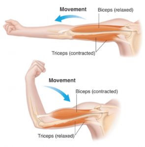
Source: Muscle movement
Anatomy of muscle fibers:
- The skeletal muscles are the most numerous. They’re made up of connective tissue-wrapped as bundles of muscle fibers.
- Many muscle bundles **(fascicles) **are bound together by fascia, a connective tissue layer, in a muscle like the biceps. There are several muscle fibers in each fascicle or a bundle of structures.
- Each muscle fiber has several myofibrilst hat run longitudinally and parallel in the sarcoplasm. Myofibrils have alternating dark and light bands that give the muscle a striated look. Myofilaments are even simpler structures that make up each myofibril.
- In a myofibril, there are two types of myofilaments. Filaments that are both thin and thick. Muscle contraction necessitates the attachment of thin and thick filaments.
Structure of Contractile Proteins:
Thin filaments, also known as actin filaments, comprise three proteins.
- It comprises two polymeric filamentous or ‘F’ actin filaments wrapped together. They’re made up of globular or G actin monomers in a polymer.
- Tropomyosin filaments are wrapped longitudinally around actin filaments.
- Troponin covers the active binding sites for myosin and is found at certain places on tropomyosin.
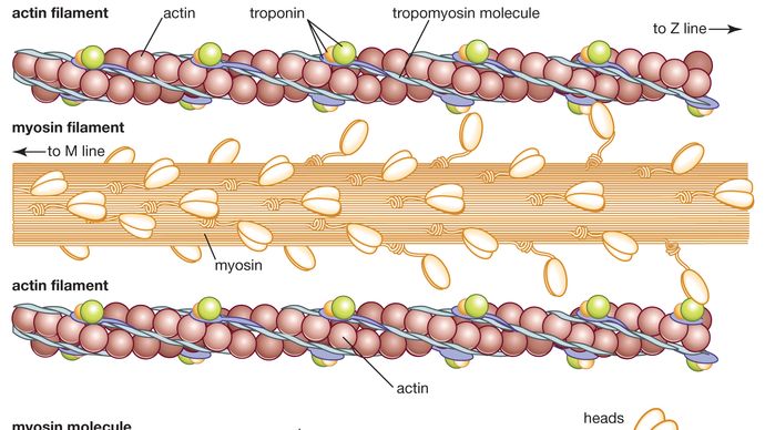
Source: Actin and myosin filaments
Myosin filaments are thick filaments made up of myosin.
- It’s a polymeric protein made up of monomeric units of the meromyosin protein.
- It comprises three components: a tail, a short arm or neck, and a spherical head.
- ATP and actin have binding sites in the globular head.
- The enzyme ATPase is found in the head.
Mechanism of Muscle contraction:
- The fundamental unit of muscular contraction is the sarcomere. It is made up of ordered repeating units of actin and myosin filaments.
- Interweaving filaments produce the ‘Z’ line that connects sarcomeres at the end. It is a sarcomere’s limiting membrane.
- The sliding of actin and myosin filaments over each other causes muscle contraction.
- Sliding causes the thick and thin filaments to overlap more and the sarcomere to shrink. As a result of this, the muscles undergo contraction.
Skeletal System:
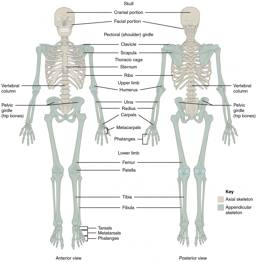
Source: Skeletal system
The skeletal system serves as our body’s structural framework and aids in movement and mobility. It shields the interior organs from harm. Bones and cartilage are two types of connective tissues that make up our skeletal system.
**Bones: **
- In an adult human person, there are 206 bones in total. Because of the calcium salts in the matrix, bones are hard, but cartilage includes **chondroitin salts. **
- The human skeleton is split into two sections: the axial skeleton, which has 80 bones, and the appendicular skeleton, which has 126 bones.
- Skull, spinal column, ribs, and sternum make up the axial skeleton (80 bones).
- The pectoral and pelvic girdle and limbs make up the appendicular skeleton (126 bones).
- The vertebral column is made up of 26 serially ordered pieces called vertebrae that are positioned dorsally. The sternum is a flat bone on the thorax’s ventral midline.
- The ribs are divided into 12 pairs. Each rib is a narrow flat bone that connects the vertebral column dorsally and the sternum ventrally. The eighth, ninth, and tenth pairs of ribs do not articulate directly with the sternum but instead use hyaline cartilage to connect to the seventh rib. They are termed as** Vertebrochondral (false) ribs. **The last two pairs of ribs (11th and 12th) are called floating ribs because they are not attached ventrally.
- The bones of the limbs along with their girdles constitute the appendicular skeleton. Each limb is made of 30 bones.
- A cup shaped bone called patella covers the knee ventrally (knee cap). Pectoral and Pelvic girdle bones help in the articulation of the upper and the lower limbs respectively with the axial skeleton.
- Below the acromion is a depression called the glenoid cavity which articulates with the head of the humerus to form the shoulder joint. This bone is commonly called the collar bone.
Joints:
Between bones and between a bone and cartilage, there are joints. Joints aid in locomotion and movement.
There are three different kinds of joints:
-
Fibrous Joints: do not allow any movement and are seen in the skull.
-
Cartilaginous Joints: have limited mobility, such as the intervertebral disc between two vertebrae in the spine.
-
Synovial Joints: Synovial Joints are movable joints. They feature a fluid-filled synovial space between two bones that allows for a lot of mobility.
Examples- Ball and socket joint (between humerus and pectoral girdle),** hinge joint** (knee joint), pivot joint (between atlas and axis).
Disorders of the muscular and skeletal system
- Myasthenia gravis: It is an autoimmune disease that affects the neuromuscular Junction. It leads to fatigue and even paralysis of skeletal muscle.
- Gout: It occurs because of the inflammation of joints that are caused by accumulation of uric acid crystals.
- Muscular dystrophy: It is a progressive degeneration disorder that occurs due to genetic disorders.
- Arthritis: Inflammation of joints is named arthritis.
- Tetany: It leads to spasms or rapid contractions in muscle due to low Ca++ in body fluid.
- Osteoporosis: It occurs with old age because of decreased bone mass. This increases the chance of fractures.