CBSE Class 11 Biology Chapter 6 Revision Notes Part 1
Chapter 6: Anatomy of Flowering Plants Revision Notes Part 1
The anatomy of a plant consists of different types of tissue: meristematic and permanent tissue. There are different types of permanent tissue, including the parenchyma, collenchyma, sclerenchyma, and vascular bundles. In order to examine plant anatomy, we can use plant histology. The flowering plants also have comprehensive anatomy, consisting of primary reproductive organs and secondary accessory organs.
The Anatomy of a Plant
Tissue type is an important aspect of plant anatomy. Plants tissues are of two types:
- The meristematic tissue: These tissues consists of cells that divide actively
- The permanent tissue: These tissues consists of cells that don’t divide further after achieving their specialized structure.
There are different types of permanent tissues:
-
Parenchyma is a simple tissue with isodiametric cells and thin cell walls made of cellulose. The function of the parenchyma cells is to carry out photosynthesis, secretion, and storage.
-
Collenchyma is a simple tissue with closely packed cells and no intercellular spaces. It also contains deposits of cellulose and pectin within the cell wall. The function of collenchyma is to provide mechanical support and to assimilate food.
-
Sclerenchyma is also a simple tissue with cells that usually have heavily thickened secondary walls containing lignin. It surrounds dead cells with no protoplasm. Its function is to provide mechanical support, and it is also present in the seeds and walls of fruits.
-
Xylem is a complex tissue of tracheids, vessels, xylem fibers, and xylem parenchyma. The vessels and tracheids are the main transporting materials. The function of the xylem vessels is to transport water from the roots to the stem and the leaves.
-
Phloem is a complex tissue. Angiosperms are made of companion cells, phloem parenchyma, sieve-tube elements, and phloem fibers. Its function is to transport food.
The anatomy of a plant also consists of tissue systems. These are:
-
The epidermal tissue system, which includes the outer covering of plants, consists of the epidermis, cuticle, stoma, and epidermal extensions. Through transpiration, the stoma controls the exchange of water. The stoma is covered by guard cells that contain chloroplasts.
-
The ground tissue system forms the tissue bulk between the epidermal and vascular tissues. It includes the cortex and the pith and consists mainly of simple tissue.
-
The vascular system consists of the complex tissue of the xylem and phloem. Dicotyledons have an open vascular bundle (xylem and phloem), whereas monocotyledons have a closed vascular bundle. Stems and leaves have a circular arrangement of vascular bundles wherein the xylem and radius are present within a radius.
-
The meristematic tissue refers to cells or group of cells that can divide.
Anatomy of Monocotyledons and Dicotyledons
1. Root
a. Dicots: Dicot plants have a taproot system wherein the outermost layer is called the epidermis. Following the epidermis is the multi-layered cortex, which is loosely made up of parenchyma cells. In dicotyledons, the pith is not at all distinct. There are also 2 to 4 xylem and phloem systems.
b. Monocots: Similar to dicot roots, except that there are a greater number of xylem systems.
2. Stem
a. Dicots: The stem is solid, consisting of the epidermis, cortex, and hypodermis. The epidermis is covered with a thin layer of cuticle and possesses a few stomata. Circular vascular bundles are present in dicots.
b. Monocots: The anatomy is similar to that of dicots, however, there are some differences — the hypodermis of the cortex in monocots is made up of sclerenchymatous cells. Additionally, the phloem parenchyma is missing.
3. Leaf
a. Dicots: In dicots, the lamina is made up of the epidermis, mesophyll and vascular systems. The epidermis is covered on the outer surface by cuticles and stomata. The chloroplasts are present in the mesophyll. Vascular bundles are surrounded by sheath cells.
b. Monocots: There is no different palisade and spongy parenchyma within the mesophyll.
Plant Histology
Histology refers to the study of tissues and cells under a microscope. Plant histology is important since it allows the close examination of the various parts of the plant, allowing the observer to note and compare plant anatomy, amongst other things.
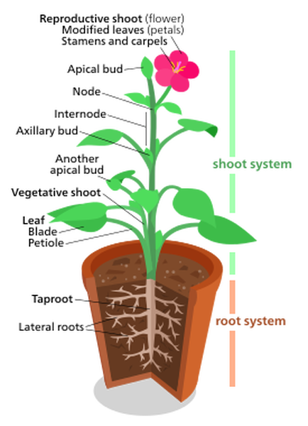
The Anatomy of a Flowering Plant (Angiosperms)
A flowering plant is also known as an angiosperm. On each flower is the floral axis upon which the essential reproductive organs (stamens and pistols) are present. On the floral axis, there are also accessory organs that help attract insects for pollination (such as sepals and petals).
There are usually four whorls of flower parts:
- A calyx made of sepals
- A corolla made of petals
- An androecium – a group of stamens
- A gynoecium that is made of pistils
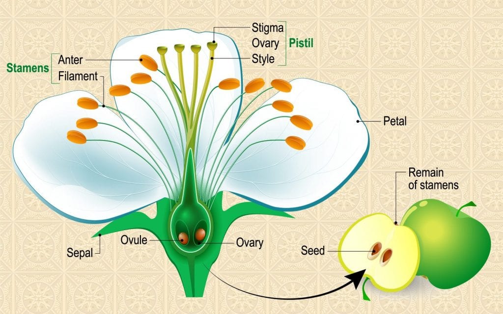
A flower that has sepals, petals, stamens, and pistils is a complete one. Usually present together, though not always, are the stamens and pistils. A flower that lacks stamens is pistillate, or female, while one that lacks pistils is said to be staminate or male.
Stamens consist of the anther and the filament. Pistils consist of the stigma, ovary, and style. The petal and the sepal envelop the stamens and pistils.
Growth in Angiosperms
The key to growth in angiosperms is the meristem tissue, which consists of undifferentiated cells that can continue to divide. Meristem allows plant stems and roots to grow longer (primary growth) and wider across the ground (secondary growth).
Sources:
Anatomy of Flowering Plants. https://ncert.nic.in/textbook.php Accessed 18 Dec, 2021.
]]>