CBSE Class 12 Biology Chapter 3 Revision Notes
Chapter 3: Human Reproduction
The Male Reproductive System

- The male reproductive system consists of a pair of testes and accessory glands, ducts, and external genitalia and is located in the pelvic area.
- The testes are positioned just outside the abdominal cavity and encased in a sac-like structure known as the scrotum.
- The rete testis, vasa differentia, epididymis, and vas deferens are the male sex accessory ducts.
- The urethra connects the urethral meatus to the outside environment.
- The penis, the male external genitalia, is enveloped with foreskin, a loose flap of skin.
- The urethra, which originates in the urine bladder and extends through the penis towards its external entrance, the urethral meatus, stores and transports sperm from the testes to the outside.
- The paired seminal vesicles, prostate, and paired bulbourethral glands are all-male accessory glands.
- The seminal plasma, which includes fructose, calcium, and enzymes, is formed by the secretion of these glands.
The Female Reproductive System
- The female reproductive system’s internal and external sex organs include a pair of ovaries and oviducts, cervix, uterus, vagina, and external genitalia located in the pelvic area.
- Along with the mammary glands, these female reproductive organs are physically and functionally united to support the whole reproductive process, comprising ovulation, fertilization, pregnancy, and childbirth.
- The oviducts(fallopian tube), vagina, and uterus make up the female accessory ducts.
- Mons pubis, labia minora, labia majora, clitoris, and hymen are the parts of the female external genitalia.
Menstrual Cycle
- For females menstruation begins at puberty and is known as menarche.
- It lasts till the female attains the age of 50 and the end of the cycle is called menopause.
- During the follicular stage of this cycle, the primary follicles in the ovary grow and mature into graafian follicles.
- Simultaneously the endometrium (lining of the uterus) regenerates through proliferation.
- The secretion of gonadotropin increases during the follicular phase. This stimulates the follicular development and also the secretion of estrogen.
- LH and FSH attain their peak in the middle of the cycle. (On the 14th day for a female who has a 28 day cycle.)
- The surge in LH induces rapture of the graafian follicle and the ovum is released.
- The endometrium is maintained by progesterone which is secreted in large amounts by the corpus luteum.
- The reproductive phase in a human female extends from menarch to menopause.
Process of Human Reproduction
GametogenesisOogenesis: is the term used to describe the process of formation of mature female gametes.
- The gametes mother cells begin to divide and enter prophase-I of meiotic division, where they are temporarily arrested and referred to as primary oocytes.
- The primary follicle has a layer of granulosa cells that surrounds each primary oocyte.
- The secondary follicle, surrounded by several layers of granulosa cells, transforms into a tertiary follicle with a fluid-filled cavity called the antrum.
- The tertiary follicles then mature into the Graafian follicle, which ruptures to release secondary oocytes (ovum) from the ovary during the ovulation process.Spermatogenesis: spermatogenesis, which begins at puberty, produces sperm in the testes.
- The spermatogonia (immature germ cells) on seminiferous tubules’ inner surface grow and increase in quantity by mitotic division.
- Spermatogonia produces spermatocytes that divide during meiosis to create secondary spermatocytes.
- Spermiogenesis is the process through which spermatids are converted into spermatozoa.
- The sperm heads stay in Sertoli cells and are discharged from seminiferous tubules during the spermiation process.
- Sperm are tiny structures with a head, neck, middle portion, and tail.
- The sperm head has an extended haploid nucleus with a cap-like structure called an acrosome covering the front half.
Fertilization
Fertilization is the process of sperm and ovum fusing.
- Semen is discharged into the vaginal canal during coitus (copulation).
- The motile sperms swim quickly to reach the isthmus and ampulla of the fallopian tube’s junction.
- The ovum also makes its way there, and gametes fuse at the ampullary-isthmic junction.
- The sperm acrosome performs an acrosomal reaction, releasing sperm lysins that break down the egg envelopes locally and open the way for sperm entry.
- These sperm lysins contain hyaluronidase, a lysing enzyme that dissolves the hyaluronic acid polymers in the intercellular spaces that hold the granulosa cells of the corona radiata together.
- It also contains corona penetrating enzyme (which dissolves the corona radiata), and acrosin, a lysing enzyme that dissolves the hyaluronic acid polymers in the intercellular (which dissolves the zona pellucida).
- The zona pellucida is then dissolved.
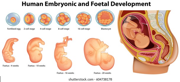
Embryonic Development
- Chorionic villi are finger-like extensions on the trophoblast after implantation.
- These along with the uterine wall, constitute a functional unit between the growing embryo and the mother body termed the placenta.
- An umbilical cord connects the placenta to the fetus, transporting nourishment and oxygen to the embryo.
- The placenta produces the hormones hCG (human chorionic gonadotropin), hPL (human placental lactogen), and relaxin in women exclusively during pregnancy.
- After implantation, the inner cell mass (embryo) divides into two layers: ectoderm on the outside and endoderm on the inside.
- Between the ectoderm and the endoderm appears a mesoderm.
- These three layers give birth to all tissues (organs) in adults.
- An embryo’s heart in a human is created after one month of pregnancy.
- Legs and fingers are established at the end of the second month.
- In 5 months, the fetus makes its first movement.
- The body is coated with fine hair by the end of 24 weeks, and eyelids and eyeless have developed.
- The fetus is completely formed at 9 months.
Parturition and Lactation
- Parturition refers to the process of delivering a fully formed fetus.
- The fully grown fetus and placenta send out parturition signals, which cause moderate uterine contractions known as the Fetal ejection reflex.
- It causes the maternal pituitary to produce oxytocin.
- Lactation is the process through which a woman’s mammary glands begin producing milk and continue to do so until the pregnancy is over.
- Colostrum is the milk produced during the first few days of breastfeeding and contains many antibodies.
Questions
- Which of these is a lytic enzyme released by the sperm? ____________
- A human primary spermatocyte has _____ autosomes.
- In a human female ovulation normally takes place ________ phase of the menstrual cycle.
- The ovum receive the sperm at the _______
- Which part of the sperm penetrates the ovum? ________
- Which of these is contained in the umbilical cord? _______
- ______ helps human sperm move.
- Cryptorchidism is a condition where one or both testes fail to descend into the scrotum. ________
- Bidder’s canal receives __________.
- Post ovulation, the mammalian egg is covered by __________.
- Which of these hormones is released from the testes? _________
- Another name for Wolffian duct ____________.
- Acrosome contains _________.
- Which of these is a temporary organ that connects a mammalian mother to its fetus? _________
- The sperm is responsible for carrying _______ to the egg.